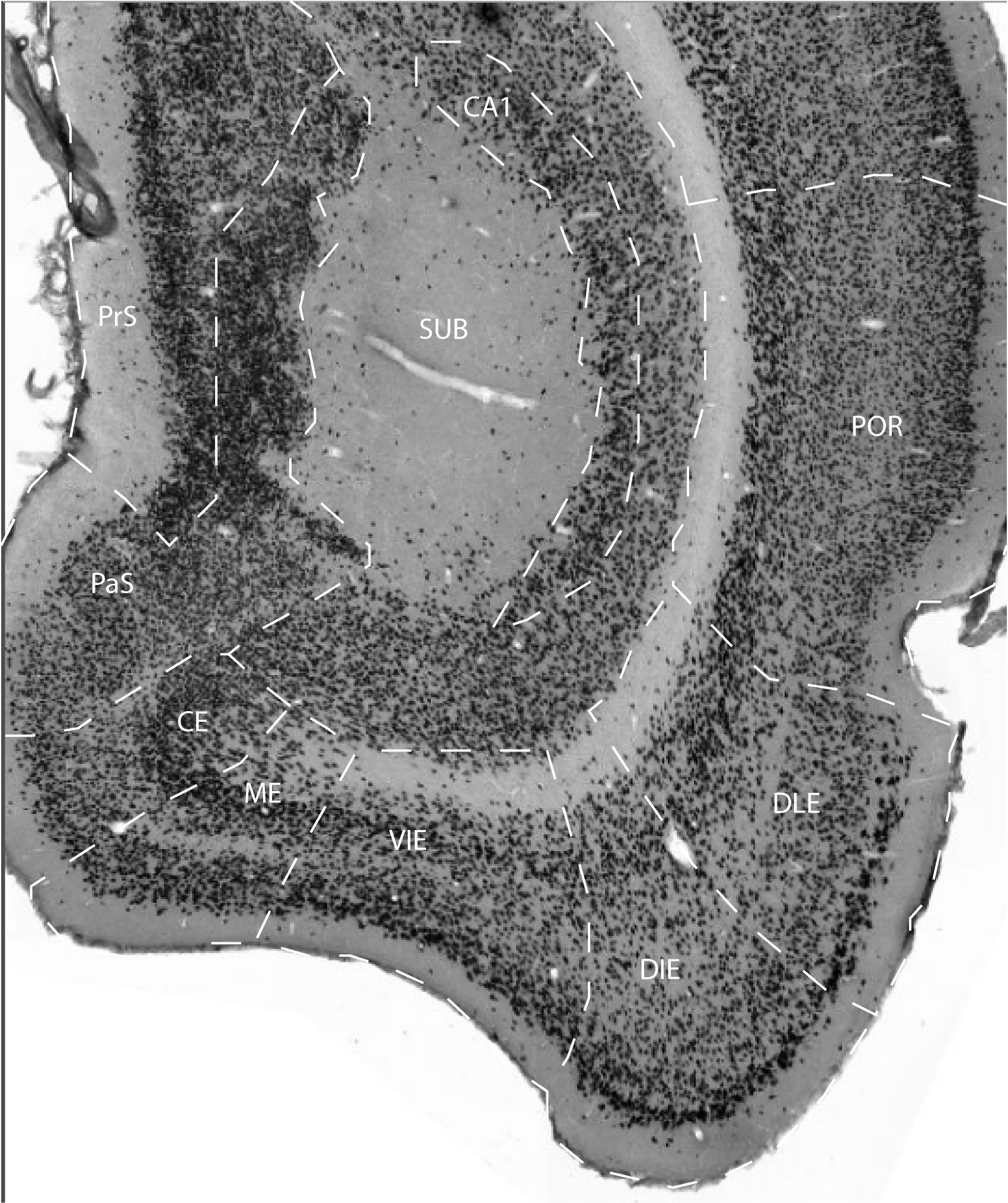
The entorhinal cortex (EC) occupies the ventro-caudal part of the cerebral hemisphere where it forms a cap-like structure. Its surface can be viewed as an ellipsoid with the white matter of the angular bundle as its center. The entorhinal cortex is hodologically defined by axonal projections from layer II neurons to the dentate gyrus of the hippocampal formation. Anteriorly, the entorhinal cortex is flanked by the piriform cortex laterally, and by the periamygdaloid cortex and the posterior cortical nucleus of the amygdala, medially (non-hippocampal regions not further described in this atlas). The transition between the entorhinal cortex and its anterior neighbors is approximately at the midst of the amygdaloid fissure, where the entorhinal cortex progressively decreases in width, such that it eventually extends anteriorly for approximately 2 mm as a narrow strip.
This anterior extension is delimited dorsolaterally by the perirhinal
cortex and ventromedially by the piriform cortex. At its laterocaudal
site, the entorhinal cortex is surrounded by the perirhinal and
postrhinal cortices. Medially, the entorhinal cortex is bordered over
most of its rostrocaudal extent by the parasubiculum. For further
cytoarchitectonic descriptions, see its 5 subdivisions; dorsal-lateral
entorhinal area (DLE), dorsal-intermediate entorhinal area (DIE),
ventral-intermediate entorhinal area (VIE), medial entorhinal area (ME)
and caudal entorhinal area (CE).
The lateral and caudal borders between EC and the perirhinal and
postrhinal cortices are characterized by:
1. the disappearance of the lamina dissecans
2. the part of EC at the border stains densely for parvalbumin whereas
the adjacent perirhinal and postrhinal cortex are almost devoid of
positive staining
3. the latter areas stain for calbindin which is much less
conspicuously present in the adjacent parts of EC
4. layer II of EC is characterized by a population of large to medium
sized neurons that stain very densely for neuronal markers such as
Nissl or NeuN. The adjacent parts of perirhinal and postrhinal cortices
are characterized by blended layers II and III consisting of small,
lightly stained neurons
The anterior border of EC is indicated by:
1. a reduction in the number of cell layers from six down to three
2. weak overall staining for parvalbumin in both anterior parts of EC
as well as the neighboring areas, so this does not provide a clear
indication of the border
3. staining for calbindin that shows less dense staining in the
anterior portions of EC compared to the adjacent regions
The medial border with the parasubiculum features:
1. a striking merge between layers II and III in the parasubiculum
2. decreased staining intensity and homogeneity for calbindin in the
neuropil of all layers
3. dense staining for acetylcholinesterase in layers I-III in
parasubiculum
References -> access a list of references
NIF Navigator (external link) -> search the Neuroscience Information Framework |
|