Area CA1 is part of the hippocampus proper, whereas the subiculum is generally considered to represent a separate entity. However, both areas share the layered composition which is typical for hippocampal fields. This layered apprearance is most easily seen in Nissl, NeuN or Timm-stained sections. In CA1, and less prominently in the subiculum, a wide molecular layer is located between the pyramidal layer and the hippocampal fissure. In the subiculum, this molecular layer can be subdivided into a deeper portion that is continuous with the stratum radiatum of CA1, and a superficial portion that is continuous with stratum lacunosum-moleculare. Note that this subdivision is difficult to detect by the markers used in this atlas. The superficial molecular layer of the subiculum and its continuation in stratum lacunosum moleculare of CA1 contain the perforant pathway fibers from the EC. Moreover, afferents from other structures such as the nucleus reuniens of the midline thalamus take a similar course and distribution.
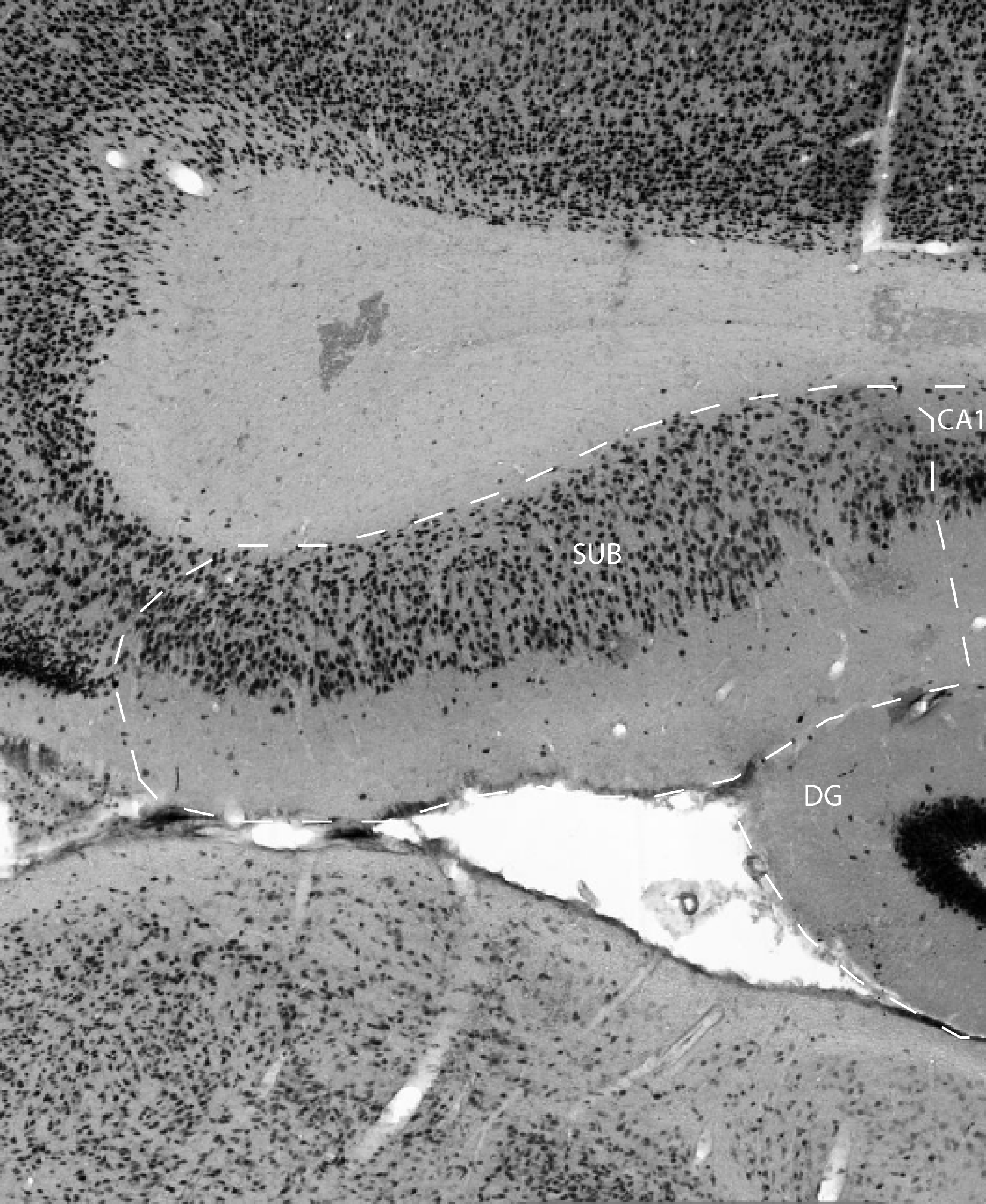
CA1 can be easily recognised in Nissl and NeuN-stained sections due to
its layer of neatly aligned pyramidal cells. This feature distinguishes
CA1 from the subiculum, which dorsally is located between area CA1 and
the retrospenial cortex, and ventrally between area CA1 and pre- and
parasubiculum. The CA1/subiculum border is clearly marked by an abrupt
widening of the pyramidal cell layer. Moreover, in material stained for
parvalbumin or AChE, the pyramidal layer of CA1 is darkly stained,
whereas the pyramidal cell layer of the subiculum is more diffusely
stained, thus indicating a marked border between the two fields. In the
TIMM staining the border is also visible, but then with an unstained
CA1pyramidal layer, and a darkly stained cell layer in the proximal
part of the subiculum. Although the border may appear easy to
establish, it is in practice not possible to determine whether a
particular dendrite at the border belongs to a neurone in CA1 or in the
subiculum. This is caused by the oblique orientation of the CA1 /
subiculum border relative to the transverse axis. Therefore, cells (in
the stratum oriens, stratum radiatum, or lacunosum moleculare) close to
this border do not necessary extent their dendrites perpendicularly to
the orientation of stratum pyramidale.
References -> access a list of references
NIF Navigator (external link) -> search the Neuroscience Information Framework |
|