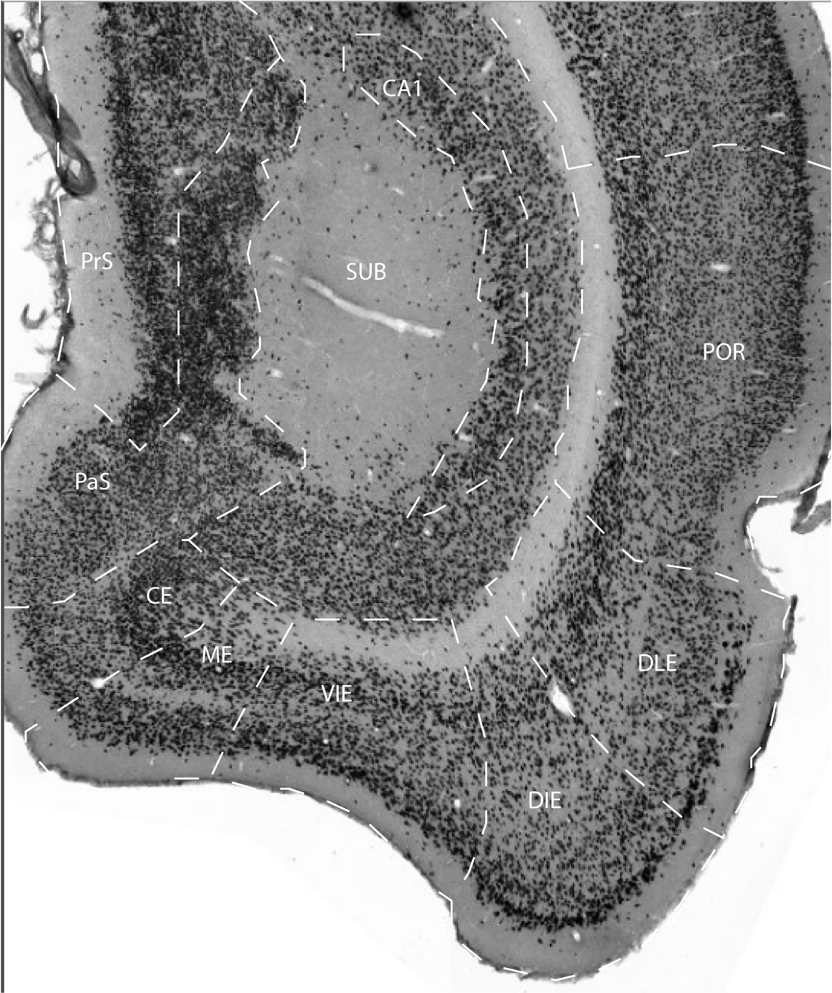
The presubiculum (PrS) and the parasubiculum (PaS) are two multilayered cortical areas positioned in between the subiculum (Sub) laterally and the entorhinal cortex (EC) or retrosplenial cortex medially. Cytoarchitectonically, PrS and PaS are alike in that, with the exception of dorsal portions of PrS, the laminar differentiation is not very conspicuous. Similar to EC, both areas have their superficial layers II and III separated from the deep layers V and VI by a prominent cell free layer or lamina dissecans. The presubiculum superficial sheet of cells consists of darkly stained small pyramidal cells. The most superficial cells, forming layer II, are the most densely packed, while the more deeper cells in layer III show a more loose arrangement. The differentiation between layers II and III becomes clearer at more dorsal levels and the cells in layer II tend to be arranged in densely packed clusters dorsally. Layer V consists of one or two rows of large pyramidal cells, and layer VI harbors a variety of neuronal types. In contrast, neurons in layers II and III of PaS are fairly large and lightly stained, and the layers are generally not differentiated from one another. The deep layers are quite similar to those in PrS and EC.The PaS, or Brodman’s area 49 has been subdivided into two portions (area 49a and 49b) (Brodman, 1909) but the subdivisions are not applied in the present account.
References -> access a list of references
NIF Navigator (external link) -> search the Neuroscience Information Framework |
|