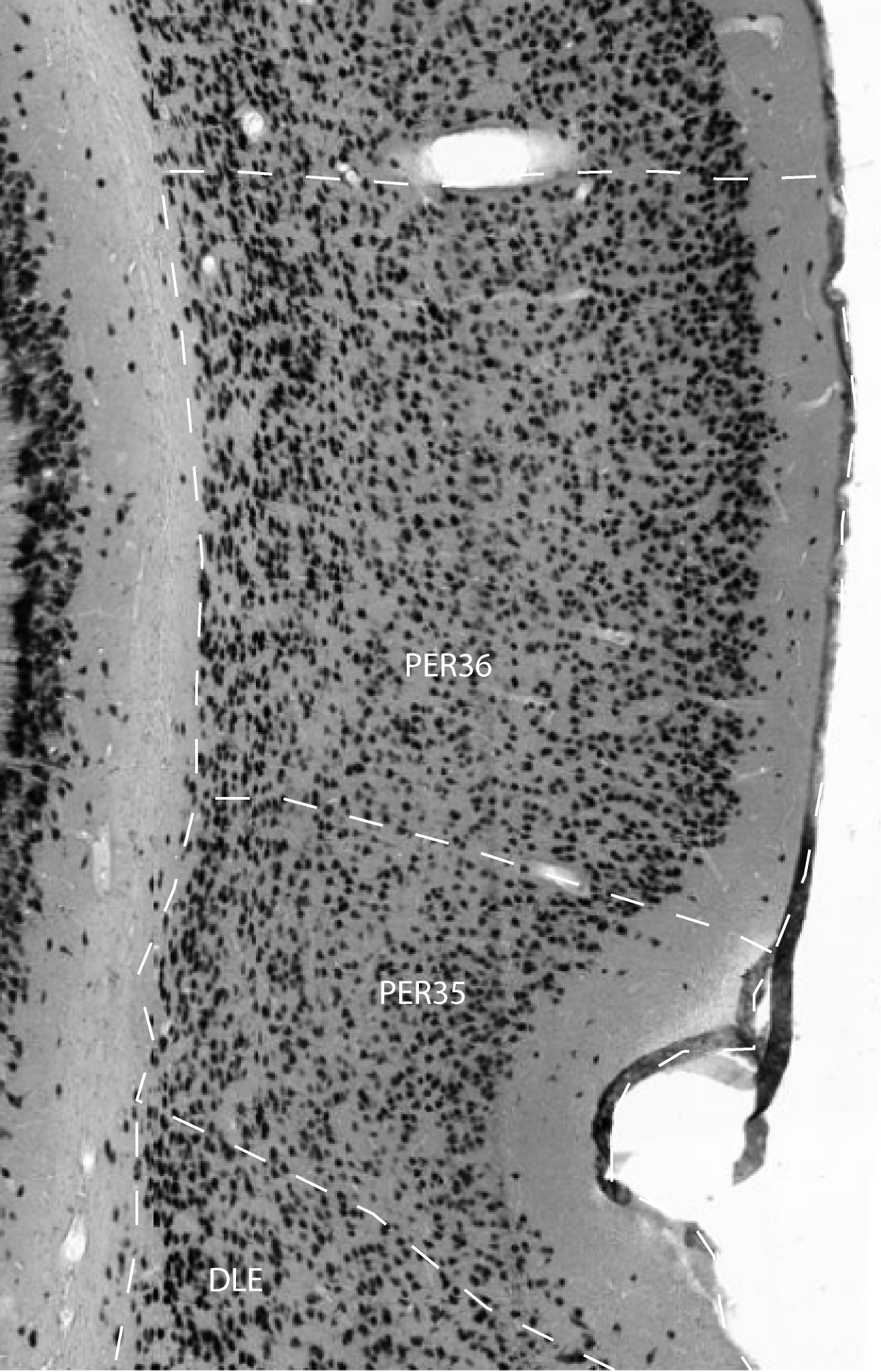
Area 35 is bordered dorsally by area 36 and ventrally and caudally by the entorhinal cortex. Just as area 36v may appear at a more anterior level than area 36d, area 35 may appear at a more anterior level than area 36. Area 35 is distinguished from the dorsally adjacent area 36 by several characteristics. First, layer I tends to be thicker, although this does not change sharply at the border between the two regions. The thickness of layer I in mid-dorsoventral area 36 is approximately 50% of that in mid-dorsoventral area 35. It may be, however, that the thickened layer I is a feature that is more appropriately associated with the rhinal sulcus than with a cytoarchitectural region. Second, the cells in area 35 exhibit a modified radial organization such that they form a shallow U-shaped arch beginning at the pial surface ventral to the rhinal sulcus and ending at the white matter deep to the rhinal sulcus. Third, area 35 is characterized by large, darkly stained, heart-shaped pyramidal cells in layer V. These cells are progressively smaller proceeding caudally. Although similar cells are seen in layer V of area 36v, there are fewer and they are not as distinctively heart shaped or as large. Heart-shaped cells become progressively smaller as one moves caudally.
Another difference is that layers II and III of area 36 are more
clearly separated into two distinct layers as compared with area 35.
Area 35 comprises two subfields, areas 35v and 35d. Area 35v sometimes
appears slightly more anteriorly than area 35d, but area 35d usually
extends farther caudally. Area 35v is distinguished from area 35d in
three ways. First, the arching organization of cells across all layers
is especially prominent in 35v, where the cells form an arch that bends
dorsally so that layer VI of area 35v merges with the layer VI of 35d.
Second, in 35d the layer II/III cells form clumps, whereas the cells in
layer II/III of area 35v are slightly elongated perpendicular to the
pial surface, giving them a "streaming" appearance. Third, in
35d the neuronal density is lower in the deep portion of layer II/III,
which gives the appearance of a cell-sparse gap between layers III and
V. This feature tends to be more evident at caudal levels.
References -> access a list of references
NIF Navigator (external link) -> search the Neuroscience Information Framework |
|