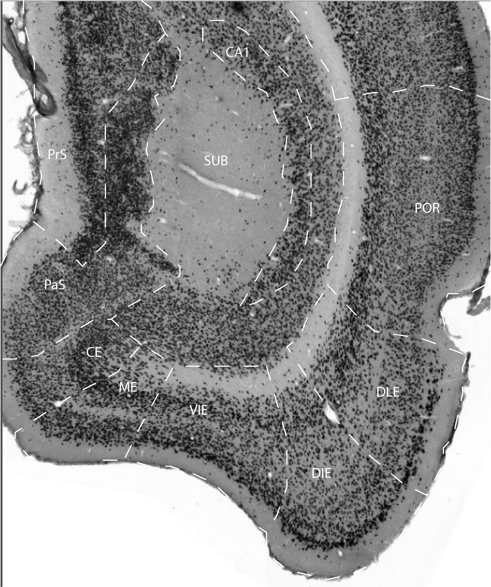
Caudally, the ventral-intermediate entorhinal area (VIE) starts at the same level, or very closely to the medial entorhinal area (ME), which borders VIE medially. The anterior border of VIE is with the amygdalo-entorhinal transitional field (AE; see below) laterally, and with the peryamygdaloid cortex, medially.
The layers of area VIE are better developed caudally than anteriorly,
where they become quite condensed and tend to merge together. Layer I
generally tends to be narrow. Layer II is also narrow and organized in
clumps that are very close to each other, rendering a continuous
appearance when studied with low magnification. Layer III is made up of
densely packed, medium-size pyramids, although not as densely packed as
in layer III of ME. Although very little or no separation exists
between layers III and V, they can still be easily differentiated since
layer V is characterized by the presence of big and dark pyramidal
cells. Such big neurons are present not only in the outer part of the
layer as in ME, but throughout the depth of layer V.
The cell density of layer V decreases at the border with layer VI,
which is rather thin and has a clear border with the underlying white
matter. The main features of field VIE are comparable to those
described by Haug (1976) for the caudomedial part of area 28L8 and the
caudal parts of VLEA as described by Krettek and Price (1977). Together
with ME, field VIE has several features in common with the rather
ill-defined, so-called intermediate entorhinal cortex of various
authors (Blackstad, 1956; Steward, 1976; Wyss, 1981; Ruth et al., 1982,
1988).
References -> access a list of references
NIF Navigator (external link) -> search the Neuroscience Information Framework |
|
