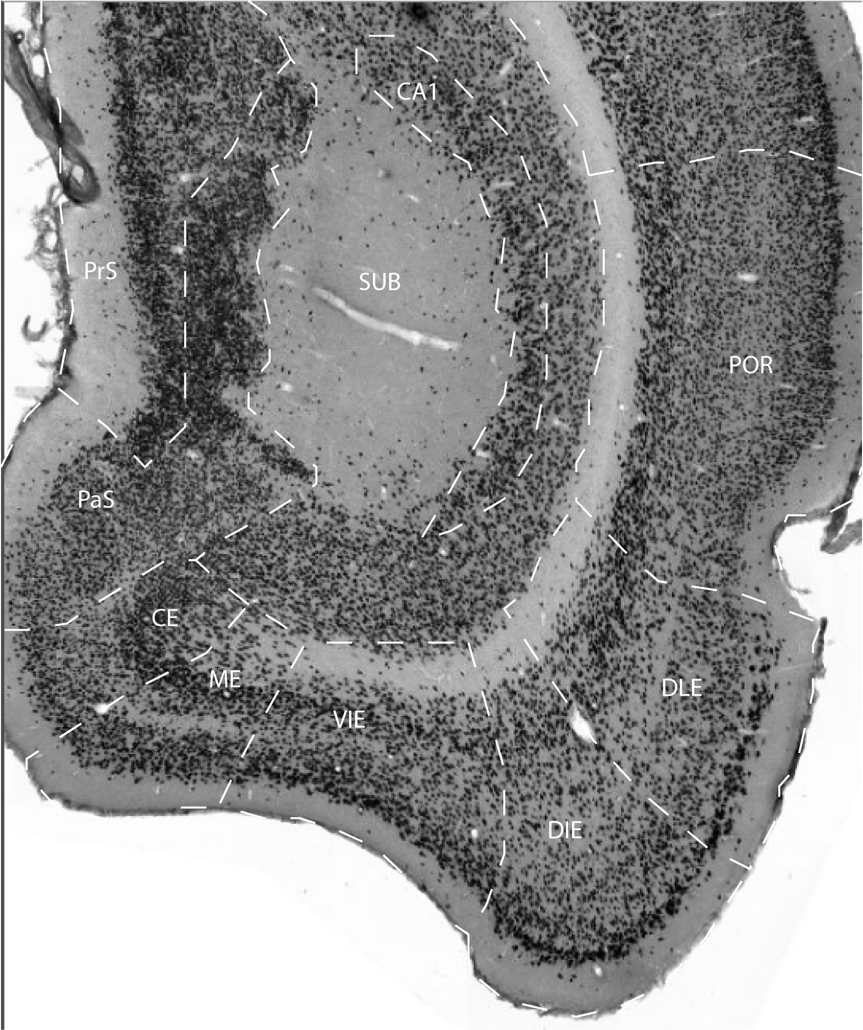
The postrhinal cortex is located caudal to area 36p and largely dorsal to the rhinal sulcus. In most cases, the postrhinal cortex arises at the caudal limit of the angular bundle when subicular cells are no longer present in coronal sections. Another landmark is the shortening of the presubiculum in the dorsoventral dimension and the imposition of a cell-sparse region deep to presubiculum that borders the underlying white matter. Like the perirhinal cortex, the postrhinal cortex is associated with the rhinal sulcus. Anteriorly, the superficial layers lie in the fundus of the rhinal sulcus, but the deep cortical layers underlying the fundus belong to the ventrally adjacent entorhinal cortex. Caudally, the region assumes a position above the fundus.
If one imagines a caudal extension of the rhinal sulcus, it would rise
at caudal levels and wrap around the caudal pole of the brain just
ventral to the postrhinal cortex. If the cortex surrounding the rhinal
sulcus and its imagined caudal extension could be straightened and
flattened, the postrhinal cortex would form a long narrow strip largely
dorsal to the sulcus and similar to the shape of the perirhinal cortex,
but shorter along the longitudinal axis. The postrhinal cortex rises
steeply and wraps obliquely around the caudal pole of the brain. Thus,
its conformation is difficult to discern in the coronal plane. Because
of the oblique cut in the coronal sections, the region extends farther
dorsally and is limited in its rostrocaudal extent. Even unfolded maps
can be misleading because of the tendency of surface areas of polar
regions to be underrepresented.
In sagittal sections, its long, narrow shape is more easily identified.
The dorsal border of the postrhinal cortex is difficult to discern but
is reliably identified relative to certain structural landmarks,
particularly the location of the parasubiculum. The parasubiculum is on
the medial cortical surface and is easily identified in cell-stained
and acetylcholinesterase-stained sections. The dorsal border of POR on
the lateral cortical surface is located directly across from the
mid-dorsoventral level of the parasubiculum. Caudally, the
parasubiculum also is useful in identifying the ventral border of the
postrhinal cortex. At its caudal limit, the parasubiculum extends
laterally more than halfway across the cortex and lying between the
postrhinal cortex and entorhinal cortex. Thus, at this level, the
postrhinal cortex has a modified triangular shape such that
parasubiculum (medially) and the entorhinal cortex (laterally) form one
side of the triangle and the pial surface and the lateral visual
association cortex (VISl) form the other two sides.
Perhaps the most characteristic cytoarchitectonic feature of the
postrhinal cortex is its homogeneous packing density across layers
II and IV and the resulting lack of a prominent laminar structure. It is
difficult to differentiate deep from superficial layers because the
layers appear to blend into one another. A second characteristic of the
region visible in coronal sections is that all layers become unusually
broad as one moves from superficial to deep. This thickening, however,
is entirely a function of the conformation of the region and the plane
of sectioning; the postrhinal cortex wraps around the caudal pole of
the brain, and at these levels the coronal plane cuts obliquely across
the radial axis of the cortex. A third characteristic of the postrhinal
cortex is also due to conformation: the surface of the ventral portion
of the region that is located dorsal to the rhinal sulcus is tightly
convex such that deeper layers are compressed. Similar to cortical
layers of gyri of the primate brain, the length of the superficial
layers is longer than the length of the deeper layers. As a result,
only a very narrow segment of layer VI is associated with the
superficial layers of ventral postrhinal cortex. Although the lack of a
prominent laminar structure is accentuated in coronal sections, this
feature is also apparent in sagittal sections. The broadening of deeper
layers, however, is not apparent in sagittal sections in which layers
I, III, V, and VI occupy approximately one-third each of the radial
extent of the cortex. The postrhinal cortex has two subfields: PORv and
PORd. PORv is located dorsal to the entorhinal cortex and caudal to
area 36p.
In the coronal plane, in rare cases, PORv emerges anterior to PORd, and
in these cases, PORv is located ventral to 36p at its most caudal
levels. In most cases, however, area 35d occupies this position. PORd
is located dorsal to PORv, but sometimes begins slightly more caudally
than PORv. Cells in PORd layer III are more heterogeneous in size,
shape, and color and are more organized and radial in appearance than
in the ventrally adjacent PORv. Layers II and III are each composed of
a homogeneous population of medium-sized, lightly stained round and
polygonal cells, but the cells are more densely packed in layer II. In
some cases, small dark pyramids are mixed into layer II. In the
dorsally adjacent visual/temporal cortex (Tev), the cellular packing
density is also higher in layer II; however, layer II and III cells of
the dorsally adjacent Tev at this level are small, round, and darkly
stained and do not have a radial appearance as in PORd. A granular
layer is distinguishable, but less so at caudal levels. Layer V of PORd
is slightly narrower than in PORv. Layer V differs from the dorsally
located Tev in that Tev layer V is more open and sparsely populated,
and the cells are larger.
There are several typical cytoarchitectonic features of PORv. Perhaps
the most distinctive is the presence of ectopic layer II cells at
anterior levels of the region near the border with entorhinal cortex.
These ectopic cells are present in all cases, but they vary in
prominence. Layer II cells are moderately large, light, and round, but
not as large as those seen in perirhinal cortex. Anteriorly, layers II
and III can be distinguished from one another because layer III cells
are less organized and less densely packed. Caudally, however, layer II
is not easily distinguished from Layer III. PORv is dysgranular at all
rostrocaudal levels, such that granule cells fill in between layers III
and V, giving an overall homogeneous appearance to the region.
Anteriorly, the width of layer V appears broader than in the dorsally
located PORd, but this may be secondary to the curvature of the cortex
at this level. Layer V is composed of small pyramid-shaped cells. Layer
VI, which is fused together with layer V, is composed of fusiform cells
and elongated pyramids that are oriented almost parallel with the
angular bundle; however, only a small portion of layer VI is associated
with PORv.
References -> access a list of references
NIF Navigator (external link) -> search the Neuroscience Information Framework |
|