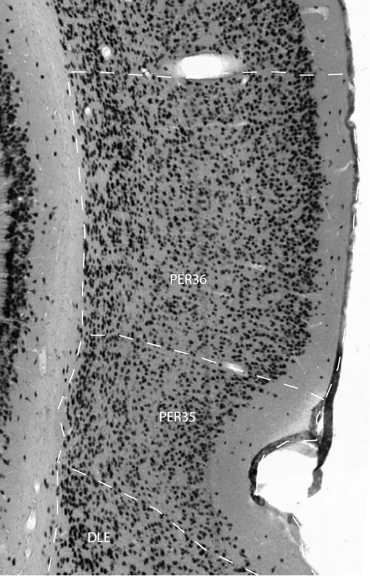
Area 36 is bordered dorsally by the ventral temporal cortex (TeV), ventrally by area 35 and caudally by the postrhinal cortex . In general, area 36 is characterized by a patchy layer II composed of aggregates of lightly-stained, medium-sized cells. Layer II of TeV also appears patchy in some animals, especially at anterior levels, but can be distinguished from area 36 because the cells are more typically pyramid-shaped and usually more lightly stained.
In area 36, granular cells are apparent, but do not form a discrete
layer rather, layer IV appears to merge with layer V. Area 36 is also
characterized by a thick, bilaminate layer VI that distinguishes it
both from the dorsally adjacent TeV and the ventrally adjacent area 35.
The outer sublayer is similar to layer V in packing density and
staining characteristics of cells. In the inner sublayer, cells are
flattened parallel to the surface of the external capsule. This
bilaminate layer VI, as well as the patchy layer II, are probably the
features of area 36 that are seen most reliably across individual
animals. Area 36 can be subdivided into three subfields: areas 36d,
36v, and 36p according to Burwell (1995).
References -> access a list of references
NIF Navigator (external link) -> search the Neuroscience Information Framework |
|