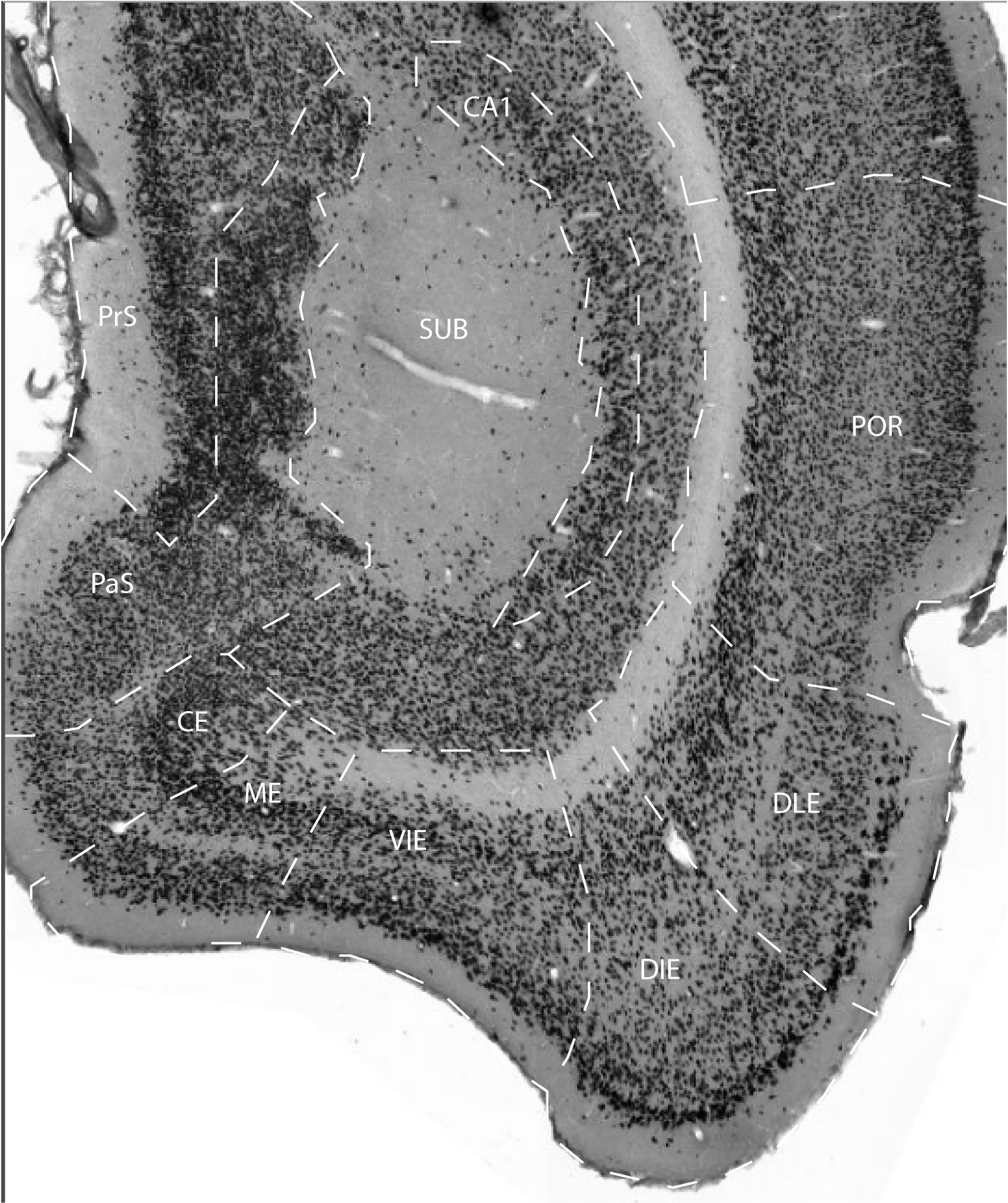
The medial entorhinal area occupies the most ventromedial portion of the entorhinal cortex. In most animals, the ME field is located on the medial bank of the amygdaloid fissure. Its overall cytoarchitectoninc features are rather similar to those of the caudal entorhinal area (CE), in that it shows a well defined laminar separation with a clear lamina dissecans, columnar organization in the deep layer V and VI, and an overall homogeneous cell density in layers II and III.
Its caudal and caudomedial border is with CE, and its medial border is
with the parasubiculum. Rostromedially, the parasubiculum is flanked by
the most anterior remnant of the presubiculum. Laterally and
anteriorly, it borders the ventral intermediate entorhinal field. A
small portion of its rostromedial border is continuous with the
periamygdaloid area.
Together with the ventral-intermediate entorhinal area (VIE), field ME
has several features in common with the rather ill-defined, so-called
intermediate entorhinal cortex of various authors (Blackstad, 1956;
Steward, 1976; Wyss, 1981; Ruth et al., 1982, 1988).
References -> access a list of references
NIF Navigator (external link) -> search the Neuroscience Information Framework |
|