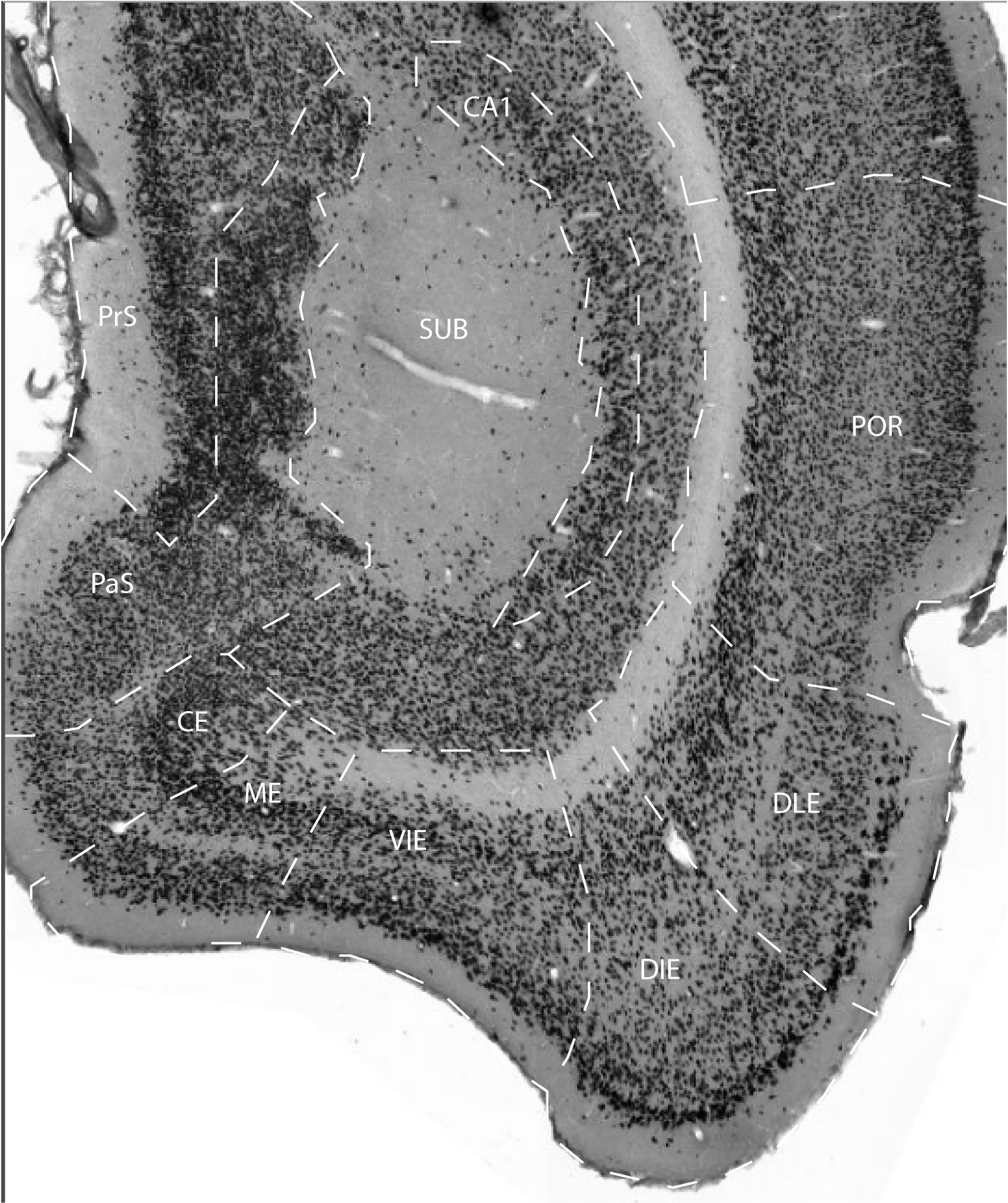
The DIE field forms the ventrolateral portion of the entorhinal cortex. Layer I is rather narrow and contains very few scattered neurons. Layer II contains rather big, rounded neurons that are distinctly stained in regular Nissl stain. A very narrow, relatively acellular band separates layer II from layer III in much of the DIE. Layer III is wide, and clearly presents a narrow, more densely packed outer zone with its neurons arranged in cell clusters, and a less densely and irregularly packed inner zone (Krettek & Price, 1977).
According to several authors (cf. Caballero-Bleda and Witter, 1993) the
outer portion of layer III of field DIE should be considered part of
layer II. This deep portion of layer II is in such instances designated
as layer IIb, whereas the more superficial portion is named IIa. Layer
IV is very poorly developed or absent, so layer III abuts on layer V.
Layer V is rather narrow and comprises loosely arranged medium- or
big-sized pyramids that are not as darkly stained as those in the ventral-intermediate entorhinal area (VIE) and the medial entorhinal area (ME). This difference is particularly striking in layer Va of these areas. Whereas layer Va is a more or less continuous row of large, darkly stained neurons in ME and to a slightly lesser extent in VIE, such cells are only incidentally present in DIE.
Layer VI is narrow, lacks a columnar arrangement, and is more compact
than layer V. This field largely coincides with the classical lateral
entorhinal cortex of Blackstad (1956; see also Steward, 1976; Wyss,
1981; Ruth, 1982, 1988), area 28L of Haug (1976), and ventromedial
portions of DLEA as described by Krettek and Price (1977).
References -> access a list of references
NIF Navigator (external link) -> search the Neuroscience Information Framework
|
|
