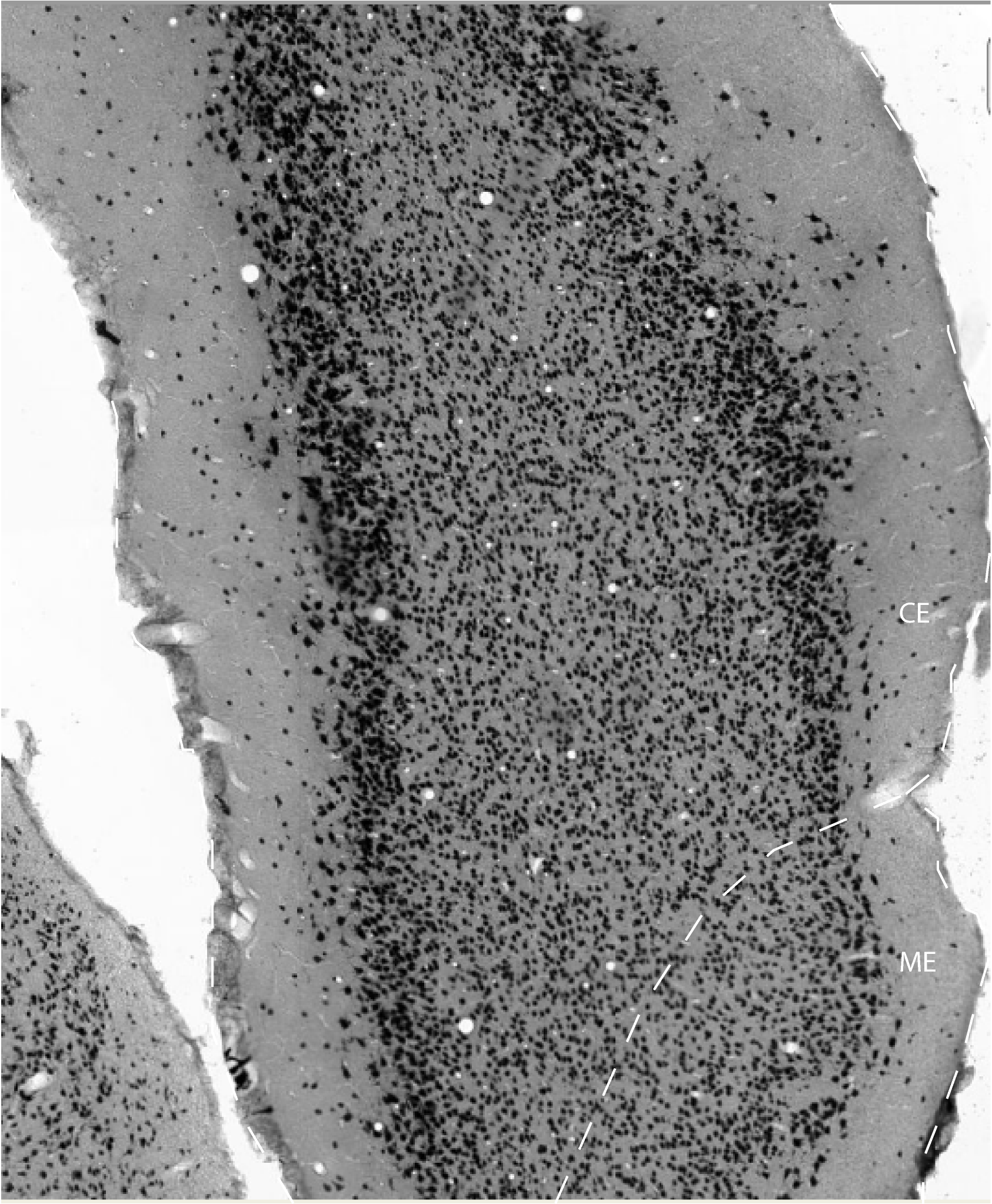
The caudal entorhinal area makes up the ventral part of the caudal pole of the rat cerebral hemisphere. At the very caudal extreme, it spans the mediolateral extent of the entorhinal cortex, from the rhinal fissure laterally to the border with the parasubiculum medially. CE has a strikingly columnar appearance. Layer II forms a continuous band of medium- or big-sized, darkly stained neurons, sharply delimited from layer I. At its medial extreme, layer II suddenly increases its width, forming a club-like thickening. Layer III contains medium-sized pyramidal cells that are homogeneously distributed. Layers III and V are separated by a clear cell free zone or lamina dissecans. Layer V is populated by medium-sized neurons that are loosely arranged at the border with the lamina dissecans, whereas in the deep part of the layer, these cells tend to be somewhat more tightly arranged. Layer VI is broad and not clearly demarcated from the white matter. In layers V and VI, neurons are oriented in a columnar fashion.
The architectonic features of the caudal entorhinal field, in
particular those of the superficial layers II and III, and the presence
of a clearly demarcated lamina dissecans are largely coincident with
the descriptions of the typical medial entorhinal cortex by previous
authors (Blackstad, 1956; Haug, 1976; Steward, 1976; Wyss, 1981; Ruth
et al., 1982). However, the extent of the medial entorhinal cortex as
defined by these authors is much larger than in our observations, and
includes the medial entorhinal field (ME).
The lateral and caudal borders between CE and the perirhinal and
postrhinal cortices is indicated by:
1. the lamina dissecans disappearing
2. the part of CE at the border stains densely for parvalbumin whereas
the adjacent perirhinal and postrhinal cortex are almost devoid of
positive staining
3. the latter areas stain for calbindin which is much less
conspicuously present in the adjacent parts of EC
4. layer II of EC is characterized by a population of large to medium
sized neurons that stain very densely for neuronal markers such as
Nissl or NeuN. The adjacent parts of perirhinal and postrhinal cortices
are characterized by blended layers II and III consisting of small,
lightly stained neurons
5. at medial levels, CE can occasionally border a laterally extending
part of PaS. This area is described below. This feature is in the rat
brain quite variable and therefore animal specific; such individual
variation is not commonly seen in rats
The anterior border of CE with ME is indicated by:
1. the rather homogeneous layer II becomes less obvious and generally
breaks into two or three clusters of cells
2. layer II is separated from layer III by a narrow, irregular cell
sparse zone
3. layer III looses its very regular density and can be subdivided in
an outer more densely packed zone and an inner less dense zone
4. the superficial part of layer V shows a population of large, darkly
stained pyramidal cells not present in CE
The anterior border of CE with the ventral-intermediate entorhinal area
(VIE) is indicated by:
1. loss of the conspicuous lamina dissecans
2. loss of the very regular appearance of layer III and the columnar
appearance of layers V and VI
3. a sudden increase in large darkly stained pyramidal cells in layer V
4. sudden thinning of layer II
The medial border with the parasubiculum features:
1. a striking merge between layers II and III in the parasubiculum
2. striking decrease in staining intensity for calbindin throughout the
neuropil of all layers
3. dense staining for acetylcholinesterase in layers I-III in
parasubiculum
References -> access a list of references
NIF Navigator (external link) -> search the Neuroscience Information Framework |
|