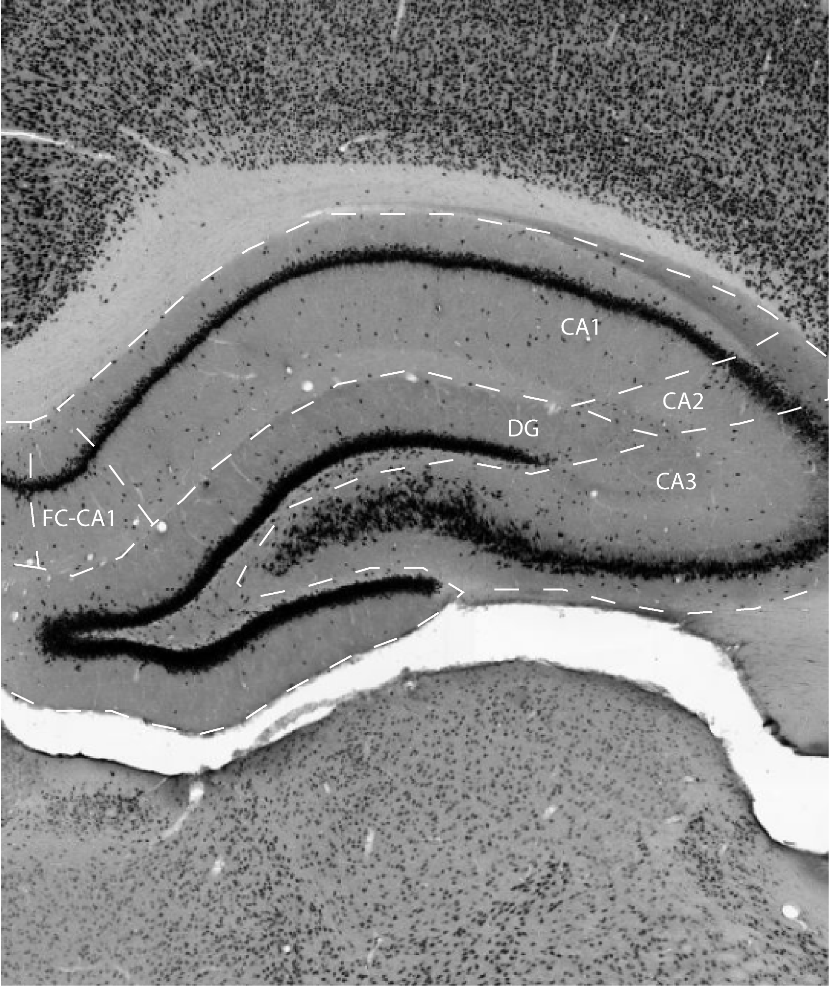
The dentate gyrus is characterized by the presence of a curved cell layer, densely packed with granule cells, i.e. cells that lacks basal dendrites but have an extensive apical dendritic tuft, extending into he molecular layer. The curved cell layer presents itself as a V-shaped, or C-shaped structure, depending on the dorsoventral level of the hippocampal formation (HF). Here, the portion of the granule cell layer adjacent to CA1, is referred to as the enclosed blade (which is synonymous with suprapyramidal, dorsal, or inner blade/limb), while the opposite portion of the granule cell layer is called the exposed blade (which is synonymous with infrapyramidal, ventral, outer, or free blade/limb).
The granule cell layer is covered by a molecular layer and it encloses
a polymorph layer, referred to as the hilus. The overall
cytoarcitecture of DG is easily appreciated in NeuN or Nissl stains.
Other staining methods do not add information to specifically delineate
DG, although with a stain for calbindin, the hilus is clearly set apart
from the adjacent proximal part of CA3 as a positive area, similar to
the stained mossy fibers that emanate from DG reaching cells in the
hilus as well as providing the main connection to CA3. This staining
pattern is mimicked by staining with an antibody against dynorphin.
References -> access a list of references
NIF Navigator (external link) -> search the Neuroscience Information Framework |
|